
Cellular Landscapes - Digital works
I studied Cellular and Molecular Biology in Academic research laboratories for 15 years.
Completely in awe of the incredible, beautiful sub-cellular structures I imaged under high-powered microscopes, I vowed to one day to turn them into art. This is some of that work.
Click here for more info or on an image to learn more about it.

As many natural forms, the 'skin', or epithelium, of the developing Drosophila embryo (vertical orientation, left side) is beautifully patterned according to ordered mathematical principles, known in some circles as 'sacred geometry' (ie. the 'flower of life', right-hand side). Three proteins comprising the 'skeleton' of the skin cells shown are labelled here in red, green and blue (overlap of red and green light looks yellow), showcasing different cell types along the length of the embryo. The vertical 'stripes' reveal the ultimate segmentation pattern of the larva that will hatch result this embryo.
Blended images: 25x magnification confocal micrograph layered with images generated in Photoshop.
(For aficionados, the blue cellular component labelled here is the cytoskeletal component spectrin; orange is cytoskeletal-associated protein, Short stop/Shot; green is acetylated tubulin)
Imaged using an Olympus Fluoview 1000 in the laboratory of my gracious mentor, Dr Nick Brown (Cambridge University), with funding from the BBSRC, and gratitude to my PhD supervisor, Dr Katja Roper (MRC-LMB, Cambridge University).

Developing musculature (teal) and central nervous system (orange) in the Drosophila embryo. Striking similarities in brain and nerve cord development between insects and vertebrates have led to many important discoveries.
25x magnification. Confocal micrograph.
(For aficionados, the blue cellular component labelled here is the cytoskeletal component actin - Rhodamine-Phalloidin stain - to label musculature; the orange is the pan-neuronal transcription factor, ElaV.)
Imaged using an Olympus Fluoview 1000 in the laboratory of my gracious mentor, Dr Nick Brown (Cambridge University), with funding from the BBSRC, and gratitude to my PhD supervisor, Dr Katja Roper (MRC-LMB, Cambridge University).

Drosophila ovarioles! (Substructures of the ovary.)
So beautiful.
I worked extensively with ovaries as a genetic model system during my PhD, in which I contributed to the characterisation of a novel protein family that is evolutionarily conserved between flies and all higher mammalian systems, including humans. I actually named the gene that represents this family! It's called "pickled eggs", or "pigs" for short, to describe the 'broken egg' phenotype which was very obvious when I looked at 'null mutant' ovaries for the first time (from flies in which I 'deleted' the gene).
The Drosophila egg chamber is a well-established model in developmental biology. Female Drosophila have two ovaries, each containing of roughly 16 ovarioles. The ovariole is a string of 6 or 7 sequentially developing egg chambers, or developing eggs; at the small end, reside 2-3 stem cells; at the large end, the mature egg. Ovarioles comprise several types of cells, including 'germline', 'somatic' and stem cells, and are a well-established model system to study cell adhesion, cell cycle regulation, cell differentiation, cell polarity, endocytosis, exocytosis, morphogenesis, cancer metastasis, developmental patterning and too many other processes to list here!
25x magnification. Confocal micrograph. Mandala effect generated in Photoshop.
(For aficionados, the blue cellular component labelled here is DAPI-stained nuclei, the pink is Eya, a transcription factors expressed in the follicular epithelial nuclei.)
Imaged using an Olympus Fluoview 1000 in the laboratory of my gracious mentor, Dr Nick Brown (Cambridge University), with funding from the BBSRC, and gratitude to my PhD supervisor, Dr Katja Roper (MRC-LMB, Cambridge University).
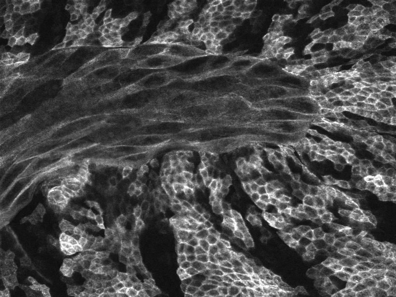
A beautiful meeting of two tissue layers - the ectoderm and the amnioserosa - in the developing Drosophila embryo.
42x magnification. Confocal micrograph.
(For aficionados, the yellow cellular component labelled here is EB1, a microtubule +-tip tracker.)
Imaged using an Olympus Fluoview 1000 in the laboratory of my gracious mentor, Dr Nick Brown (Cambridge University), with funding from the BBSRC, and gratitude to my PhD supervisor, Dr Katja Roper (MRC-LMB, Cambridge University).
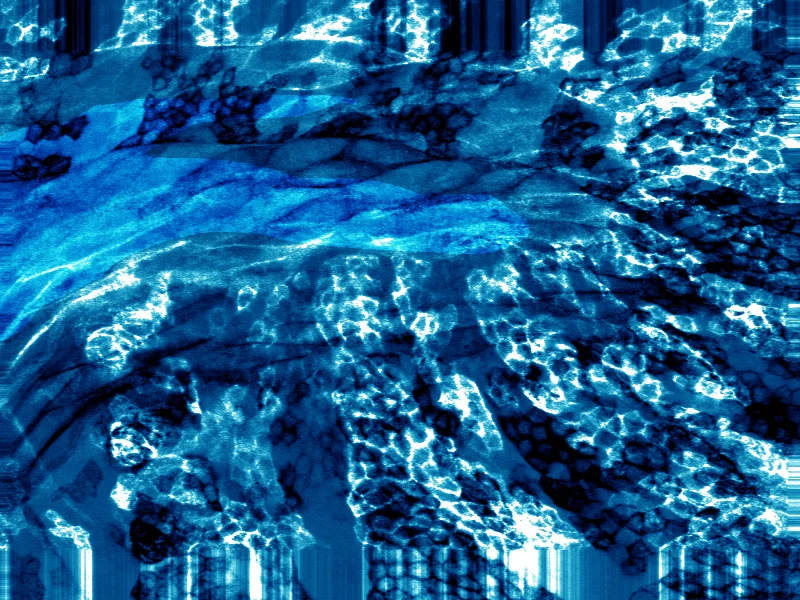
A beautiful meeting of two tissue layers - the ectoderm and the amnioserosa - in the developing Drosophila embryo.
42x magnification. Confocal micrograph.
(For aficionados, the yellow cellular component labelled here is EB1, a microtubule +-tip tracker.)
Imaged using an Olympus Fluoview 1000 in the laboratory of my gracious mentor, Dr Nick Brown (Cambridge University), with funding from the BBSRC, and gratitude to my PhD supervisor, Dr Katja Roper (MRC-LMB, Cambridge University).

Skin cells in the developing Drosophila embryo. The cellular component labelled here is tubulin, a component of the 'skeleton' of each cell that gives it shape and stability.
63x magnification. Confocal micrograph. Mandala effect generated in Photoshop.
(For aficionados, the cellular component labelled here is acetylated tubulin.)
Imaged using an Olympus Fluoview 1000 in the laboratory of my gracious mentor, Dr Nick Brown (Cambridge University), with funding from the BBSRC, and gratitude to my PhD supervisor, Dr Katja Roper (MRC-LMB, Cambridge University).
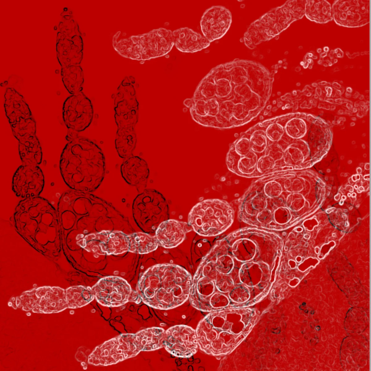
I studied cellular processes extensively using ovarioles as a model system during my PhD, in which I contributed to the characterisation of a novel protein family that is evolutionarily conserved between flies and all higher mammalian systems, including humans. I actually got to name the gene that represents this family! It's called "pickled eggs", or "pigs" for short, to describe the 'broken egg' phenotype which was very obvious when I looked at 'null mutant' ovaries for the first time (from flies in which I 'deleted' the gene).
The Drosophila egg chamber is a well-established model in developmental biology. Female Drosophila have two ovaries, each containing of roughly 16 ovarioles. The ovariole is a string of 6 or 7 sequentially developing egg chambers, or developing eggs; at the small end, reside 2-3 stem cells; at the large end, the mature egg. Ovarioles comprise several types of cells, including 'germline', 'somatic' and stem cells, and are a well-established model system to study cell adhesion, cell cycle regulation, cell differentiation, cell polarity, endocytosis, exocytosis, morphogenesis, cancer metastasis, developmental patterning and too many other processes to list here!
40x magnification. Confocal micrograph.
(For aficionados, the yellow cellular component labelled here is armadillo, a cell adhesion component.)
Imaged using an Olympus Fluoview 1000 in the laboratory of my gracious mentor, Dr Nick Brown (Cambridge University), with funding from the BBSRC, and gratitude to my PhD supervisor, Dr Katja Roper (MRC-LMB, Cambridge University).
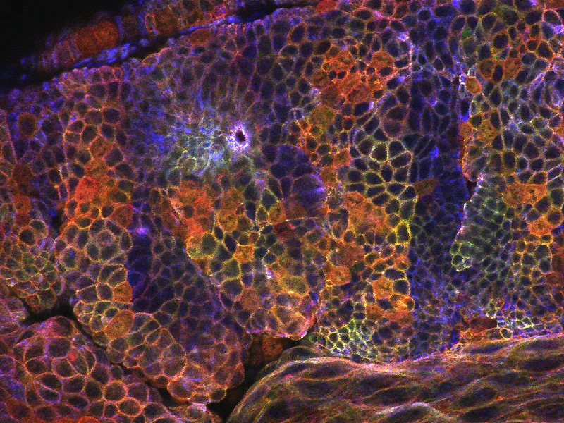
A beautiful meeting of two tissue layers - the ectoderm and the amnioserosa - in the developing Drosophila embryo.
42x magnification. Confocal micrograph.
(For aficionados, the yellow cellular component labelled here is EB1, a microtubule +-tip tracker.)
Imaged using an Olympus Fluoview 1000 in the laboratory of my gracious mentor, Dr Nick Brown (Cambridge University), with funding from the BBSRC, and gratitude to my PhD supervisor, Dr Katja Roper (MRC-LMB, Cambridge University).
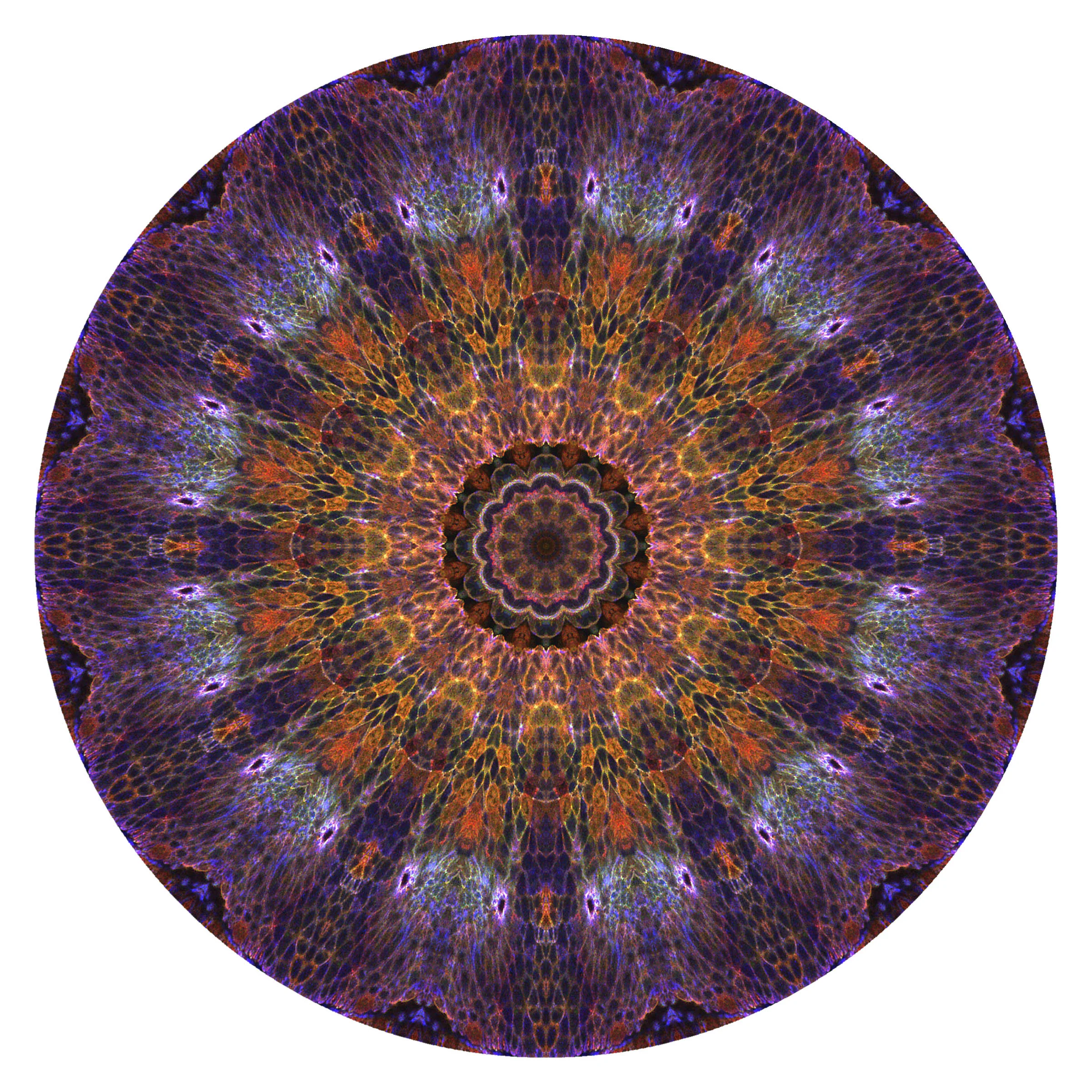
Mandala generated from Rainbow Epithelia:
A beautiful meeting of two tissue layers - the ectoderm and the amnioserosa - in the developing Drosophila embryo.
42x magnification. Confocal micrograph. Mandala effect generated in Photoshop.
(For aficionados, the yellow cellular component labelled here is EB1, a microtubule +-tip tracker.)
Imaged using an Olympus Fluoview 1000 in the laboratory of my gracious mentor, Dr Nick Brown (Cambridge University), with funding from the BBSRC, and gratitude to my PhD supervisor, Dr Katja Roper (MRC-LMB, Cambridge University).
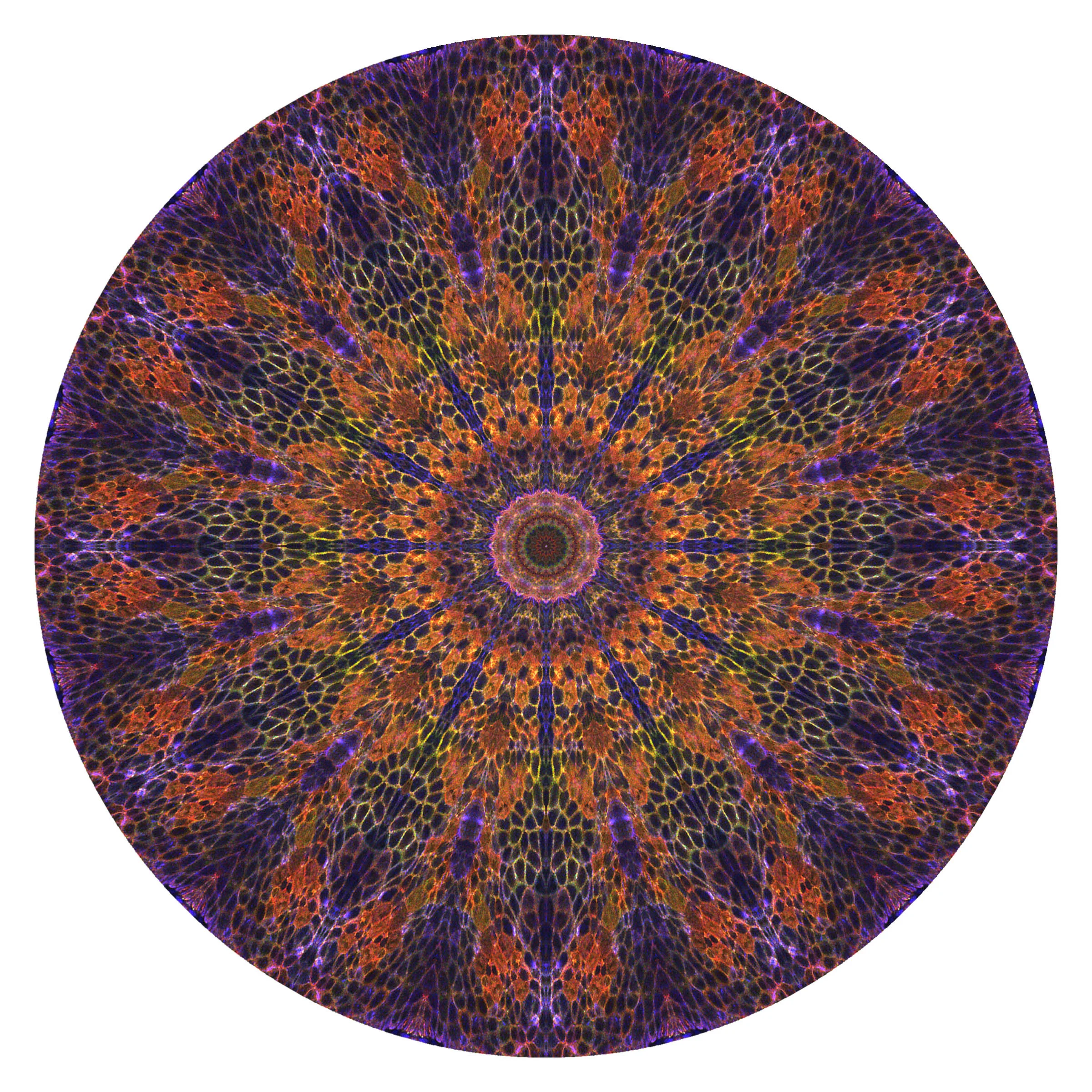
Mandala generated from Rainbow Epithelia:
A beautiful meeting of two tissue layers - the ectoderm and the amnioserosa - in the developing Drosophila embryo.
42x magnification. Confocal micrograph. Mandala effect generated in Photoshop.
(For aficionados, the yellow cellular component labelled here is EB1, a microtubule +-tip tracker.)
Imaged using an Olympus Fluoview 1000 in the laboratory of my gracious mentor, Dr Nick Brown (Cambridge University), with funding from the BBSRC, and gratitude to my PhD supervisor, Dr Katja Roper (MRC-LMB, Cambridge University).

Surface view of the Drosophila compound eye. An island of discovery with respect to visual processing, neural connectivity, cell differentiation and sorting, the fruit fly eye is one of the most advanced among insects.
42x magnification. Scanning electron micrograph.
(For aficionados, the yellow cellular component labelled here is EB1, a microtubule +-tip tracker.)
Imaged using an Olympus Fluoview 1000 in the laboratory of my gracious mentor, Dr Nick Brown (Cambridge University), with funding from the BBSRC, and gratitude to my PhD supervisor, Dr Katja Roper (MRC-LMB, Cambridge University).
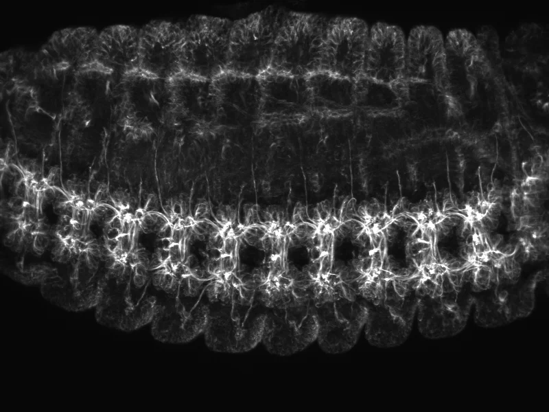
Axons from neurons of the central nervous system coalesce to form "commissures" (ladder-like structures) along the ventral midline in the fly embryo, seen here on its side. These axons forming the ventral nerve cord (analagous to the vertebrate spinal cord). Striking similarities in brain and nerve cord development between insects and vertebrates have led to many important discoveries.
42x magnification. Confocal micrograph.
(For aficionados, the cellular component labelled here is the cytoskeletal component actin - Rhodamine-Phalloidin stain.)
Imaged using an Olympus Fluoview 1000 in the laboratory of my gracious mentor, Dr Nick Brown (Cambridge University), with funding from the BBSRC, and gratitude to my PhD supervisor, Dr Katja Roper (MRC-LMB, Cambridge University).

Generated from The Developing Nervous System:
Axons of the central nervous system coalesce to form "commissures" along the ventral midline in the fly embryo (seen here on its side), forming the ventral nerve cord (analagous to the vertebrate spinal cord). Striking similarities in brain and nerve cord development between insects and vertebrates have led to many important discoveries.
42x magnification. Confocal micrograph. Starburst effect generated in Photoshop.
(For aficionados, the cellular component labelled here is the cytoskeletal component actin - Rhodamine-Phalloidin stain.)
Imaged using an Olympus Fluoview 1000 in the laboratory of my gracious mentor, Dr Nick Brown (Cambridge University), with funding from the BBSRC, and gratitude to my PhD supervisor, Dr Katja Roper (MRC-LMB, Cambridge University).
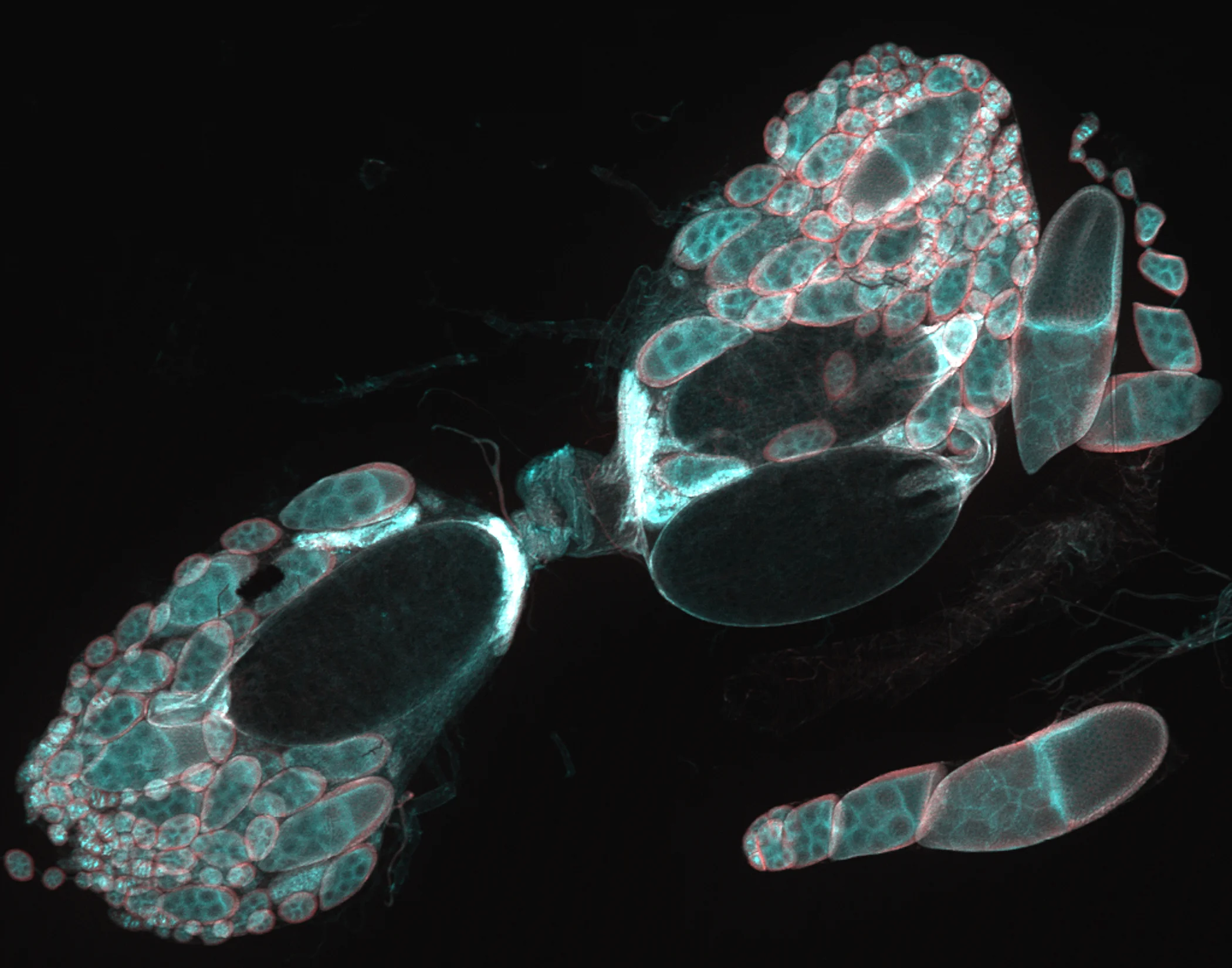
Drosophila ovaries!
So beautiful.
I worked extensively with ovaries as a genetic model system during my PhD, in which I contributed to the characterisation of a novel protein family that is evolutionarily conserved between flies and all higher mammalian systems, including humans. I actually got to name the gene that represents this family! It's called "pickled eggs", or "pigs" for short, to describe the 'broken egg' phenotype which was very obvious when I looked at 'null mutant' ovaries for the first time (from flies in which I 'deleted' the gene).
The Drosophila egg chamber is a well-established model in developmental biology. Female Drosophila have two ovaries, each containing of roughly 16 ovarioles. The ovariole is a string of 6 or 7 sequentially developing egg chambers, or developing eggs; at the small end, reside 2-3 stem cells; at the large end, the mature egg. Ovarioles comprise several types of cells, including 'germline', 'somatic' and stem cells, and are a well-established model system to study cell adhesion, cell cycle regulation, cell differentiation, cell polarity, endocytosis, exocytosis, morphogenesis, cancer metastasis, developmental patterning and too many other processes to list here!
10x magnification. Confocal micrograph.
(For aficionados, the yellow cellular component labelled here is EB1, a microtubule +-tip tracker.)
Imaged using an Olympus Fluoview 1000 in the laboratory of my gracious mentor, Dr Nick Brown (Cambridge University), with funding from the BBSRC, and gratitude to my PhD supervisor, Dr Katja Roper (MRC-LMB, Cambridge University).

A fusion of four developing Drosophila embryos (stacked vertically). Their beautiful patterning always reminds me of snakeskin so I made this 'embryonic snake' in Photoshop.
The 'skin', or epithelium, of the embryo is patterned according to ordered mathematical principles, known in some circles as 'sacred geometry'. Proteins comprising the 'skeleton' of the skin cells shown are labelled here in green and blue, showcasing different cell types and patterns in the embryo. The vertical 'stripes' reveal the segmentation patterns of the larvae that will ultimately hatch from these embryos.
25x magnification. Confocal micrographs layered in Photoshop.
(For aficionados, the green cellular component labelled here is transgenically-expresssed cytoskeletal-associated protein, Short stop/Shot; blue is acetylated tubulin.)
Imaged using an Olympus Fluoview 1000 in the laboratory of my gracious mentor, Dr Nick Brown (Cambridge University), with funding from the BBSRC, and gratitude to my PhD supervisor, Dr Katja Roper (MRC-LMB, Cambridge University).
This is mandala I created from this original image of "neuromuscular junctions" (NMJs) of motor neurons, places in the body where the axons of neurons (originating in the brain) meet muscle to form a synapse. Each blob in image is a synapse and they differ in kind. Messages from the brain that cause the muscle to contract and relax. The NMJ is only one example of many connections made between nerves and other parts of the body that result in a successfully functioning organism.
Neurons, or nerve cells, are specially designed cells that communicate using a variety of chemicals called neurotransmitters. Depending on the type of cell, specific neurotransmitters are designed to stimulate a response, assuming that a receptor is present.
At this junction, the motor neuron (labelled green here) meets muscle tissue at an axon terminal (magenta). The surface of the muscle fibre forms small ridged folds in which the end of axon to rests. Inside these folds are depressions with acetylcholine or glutamate receptors. The neuron forms synaptic vesicles that are filled with acetylcholine, glutamate or other neurotransmitters/peptides. They resemble small bulbs that will release the neurotransmitter when the muscle needs to contract.
The Drosophila larval neuromuscular system is relatively simple and its NMJs have been studied extensively as models for development and disease. They exhibit developmental and functional plasticity while displaying stereotyped connectivity, meaning that aberrations in their morphology can be used as a proxy for dysfunction. Since Drosophila NMJs are homologous to NMJs in the mammalian brain, these features make the Drosophila neuromuscular system an excellent genetic model for the study of the mammalian central nervous system.
My most recent work as a Research Associate uncovered what we believe to be a novel molecular mechanism underlying autism spectrum disorders (ASD) and schizophrenia (SCZ). We modelled the function of a family of presynaptic receptor that is essential for proper brain development and function in both flies and humans. Genetic mutations in these genes are frequently associated with ASD and SCZ and our data show potential for novel therapeutic approaches (paper in submission).
25x magnification. Confocal micrograph. Mandala effect generated in Photoshop.
(For aficionados, the green cellular component labelled here is a pan-neuronal marker (HRP), the pink is Discs Large (Dlg), a post-synaptic component.)
Imaged using a Leica TCS SP5 confocal laser scanning microscope, and with gratitude to my supervisor, Dr Michael Gordon (Dept of Zoology, University of British Columbia).